X-ray Computed Tomography scanners have been extensively used in research laboratories around the world for reservoir rock characterization and fluid flow visualization.
The scanner became a useful tool in the petroleum industry. The value of a CT lies in its ability to look inside the rock to determine flow patterns, porosity, and saturation without altering the fluids or rocks.
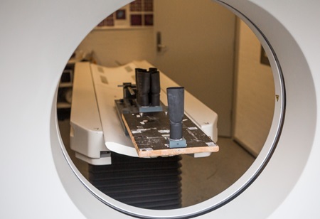
A CT-scanner is presently available in CERE. It is a fourth generation Siemens Somatom Plus with the possibility for spiral scanning and 3D image reconstruction. It has 1200 stationary detectors. The X-ray tube rotates around the object in a 360° círcular path.
The CT scanner is mainly used for our experimental investigation of the processes occurring during laboratory studies of flow in porous media of the petroleum reservoirs.
The scanner can be adjusted specifically for these experiments. A special high precision mechanism has been designed in order to precisely position the samples and to be able to repeat many scans at the same location.
A special setup has been designed in order to carry out the experiments with petroleum fluids and rock samples. It consists of an aluminum core holder (transparent for X-rays) and pumps with pressure transmitters.

The scanner is a useful instrument in many applications and has twice been used to investigate mummies first in 2000 and again in 2003.
The purpose of the first scanning was to reveal the sex of a mummy and it was published in the Danish newspaper "Politikken". The second scanning was made in collaboration with Danish Television "DR" and The Antropological Laboratory, Panum Institute, to reconstruct the face of a 2000-year-old Roman mummy. This program "Viden om" has been broadcasted on Danish and Swedish television. The scanner has also been used in food applications to measure salt distribution in pork.
Fundamental to this CT imaging technology is the ability of X-rays to pass through almost all matter and the process of mathematically reconstructing the X-ray projection data into an image. The basic quantity measured in each pixel of a CT image is the linear attenuation coefficient.
One CT scan produces an image of a slice of finite thickness of the object known as a tomogram. This slice consists of a matrix of small volume elements called voxels. After completion of a scan, each voxel is assigned a CT number which is proportional to its X-ray attenuation coefficient. The CT numbers can then be assigned gray scales or pseudo-colors and displayed as images on the monitor.
From CT images of rocks we can see if rocks contain layering, mineralogical deposits or fractures and after that determine whether rocks are homogeneous or heterogeneous. In the petroleum industry, CT-scanners have been used in experiments to determine the porosity, the permeability distribution, and the dispersion.
These are all initial tests before any core flooding experiments which include two-phase or three-phase displacements experiments are conducted. A typical experimental set-up for core flood at CERE is shown in the picture above.